Celiac Artery Compression Syndrome Radiology
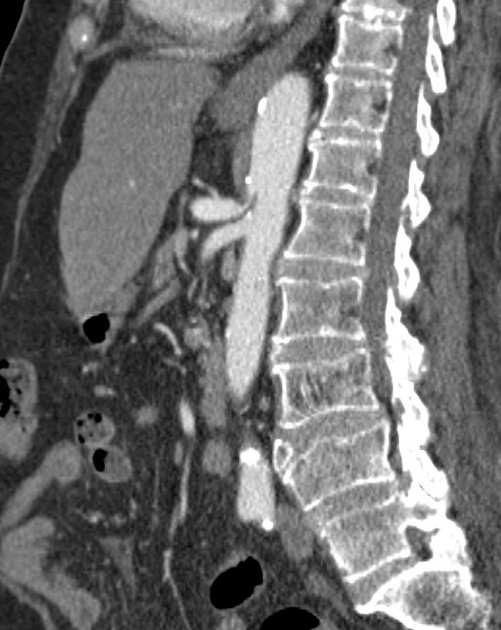
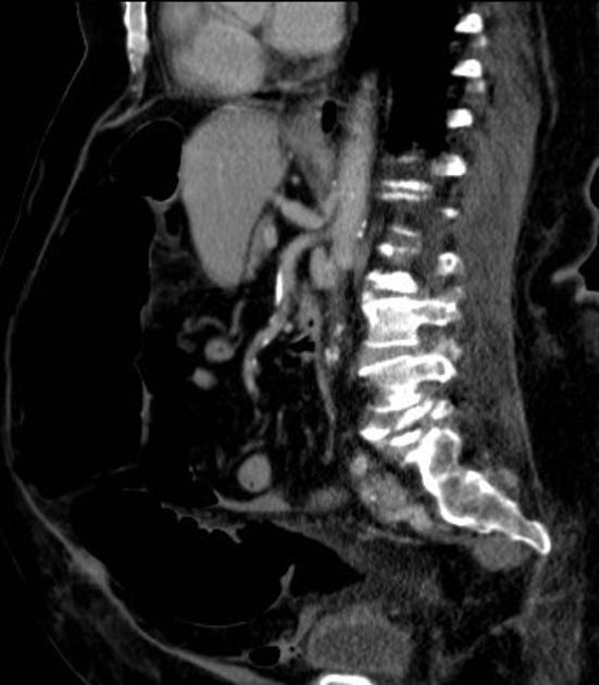
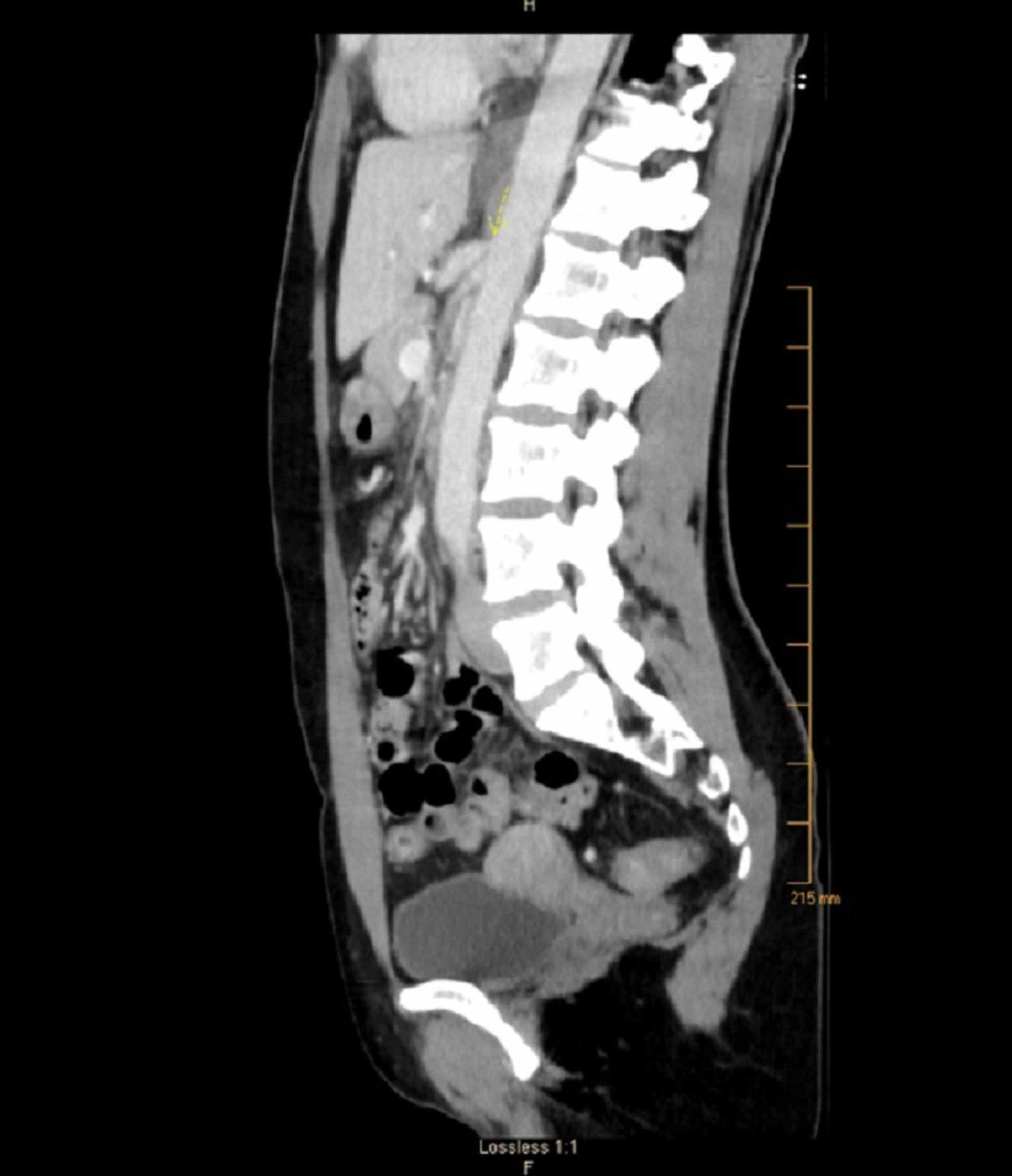
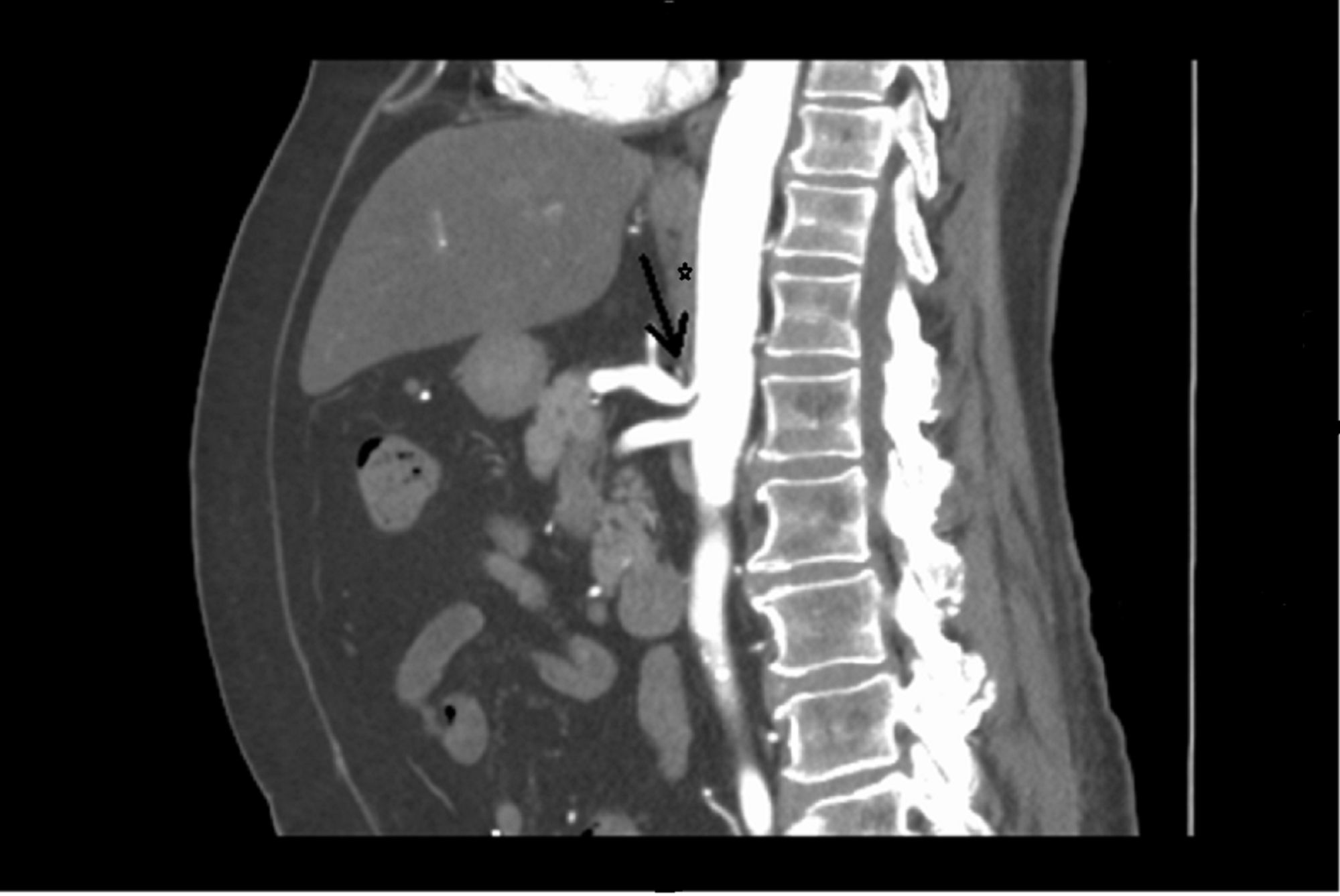
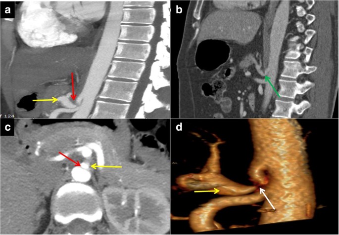
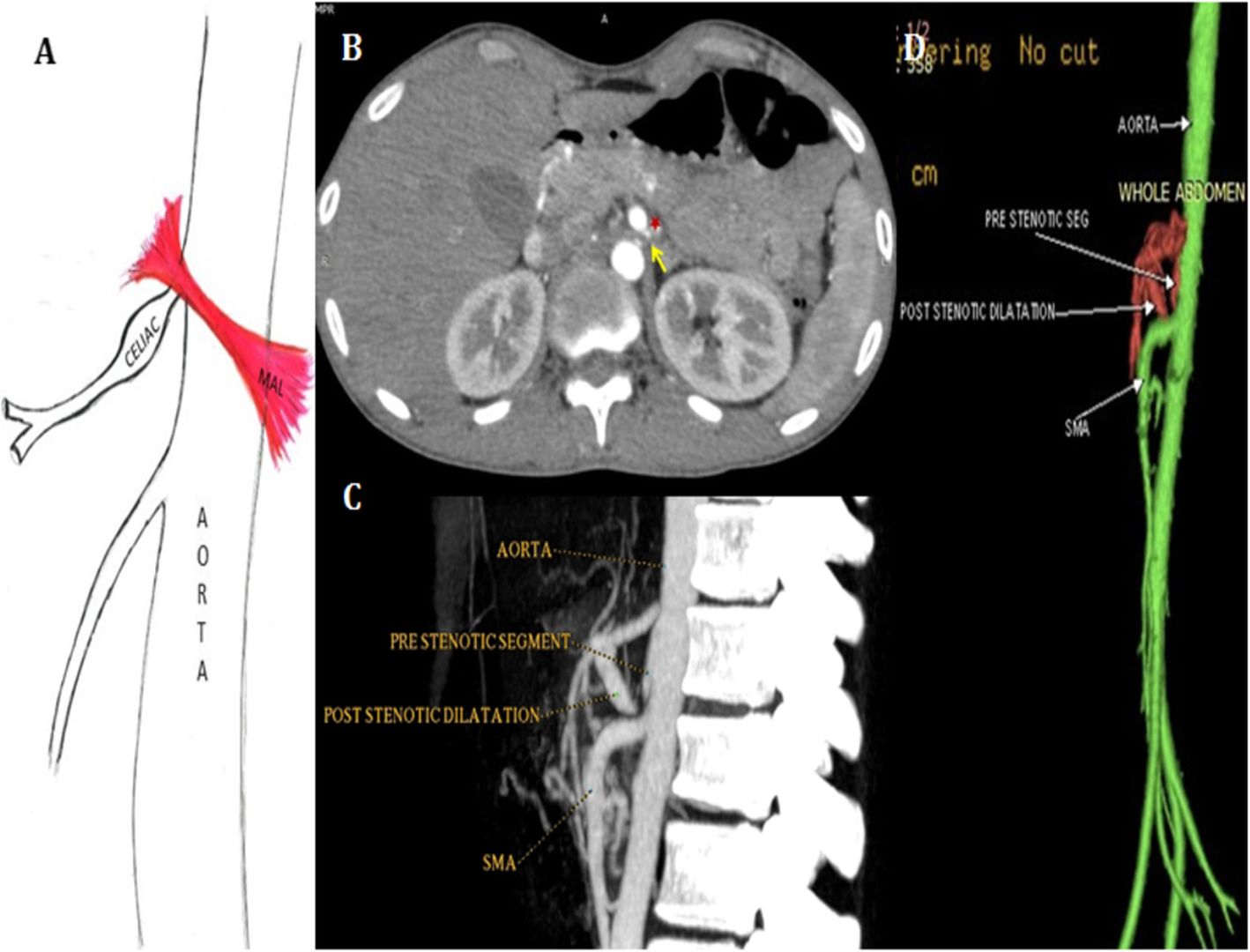
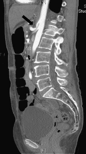
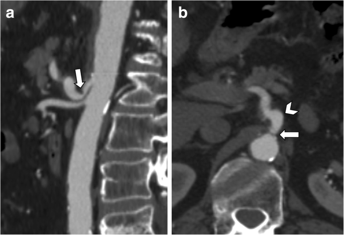
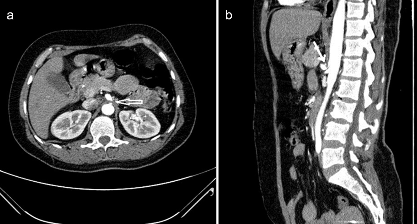
Celiac artery compression syndrome radiology. Celiac artery compression syndrome in children adolescents and young adults. Median arcuate ligament syndrome MALS also called Dunbar syndrome represents a condition characterized by external compression of celiac artery root by the median arcuate ligament Fig. Clinical and color duplex sonographic features in a series of 59 cases.
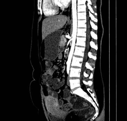
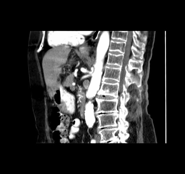
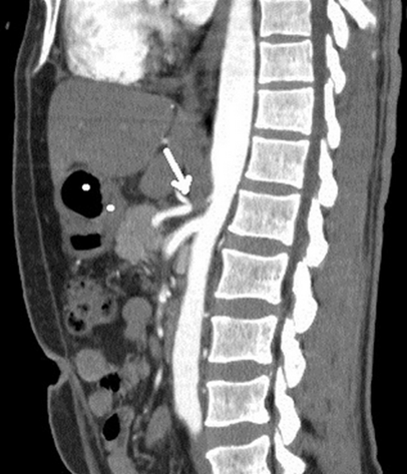
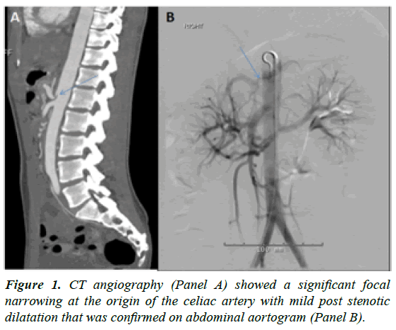
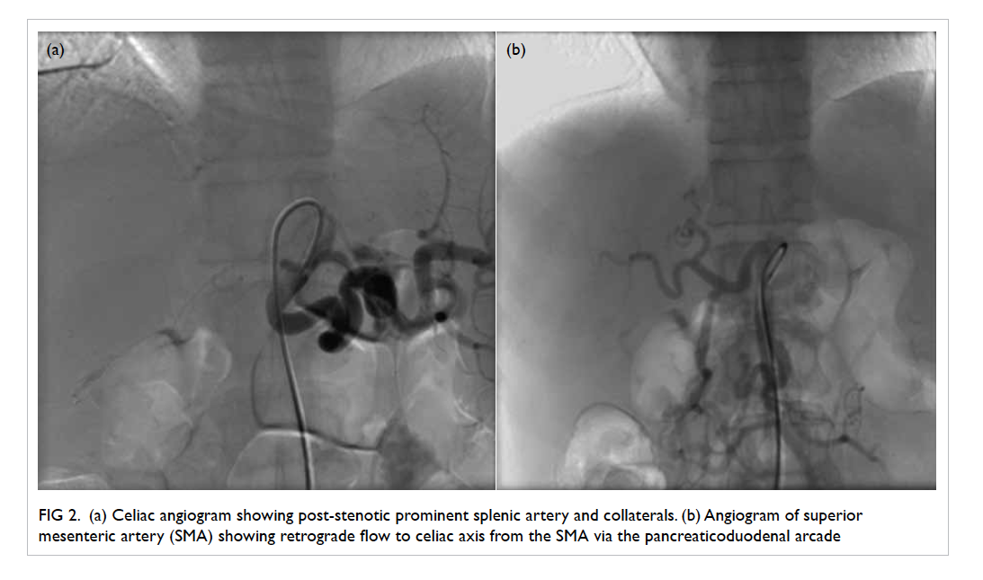
Ischemia results from external compression of the proximal celiac artery during respiration typically during expiration by the median arcuate ligament just below the diaphragm. However the condition can now be diagnosed with three-dimensional computed tomographic CT angiography. Keywords Key words celiac artery celiac artery compression syndrome CTA extrinsic compression.
Median arcuate ligament syndrome MALS is a rare disorder characterized by chronic recurrent abdominal pain related to compression of the celiac artery which supplies blood to the upper abdominal organs by the median arcuate ligament a muscular fibrous band of the diaphragm. Median arcuate ligament MAL syndrome MALS also known as celiac artery CA compression syndrome and Dunbar syndrome occurs because of extraluminal compression of the CA root by the MAL which is part of the diaphragm. Celiac artery compression syndrome is a rare condition with a reported incidence of 2 per 100000 population.
The compression of the proximal part of the celiac trunk by median arcuate ligament of the diaphragm during expiration is defined as median arcuate ligament syndrome. This clinical condition known as celiac artery compression syndrome CACS has proven controversial in definition and relevance. Celiac artery compression syndrome also known as median arcuate ligament syndrome is a condition where a muscular fibrous band of the diaphragm the median arcuate ligament compresses the celiac axis which supplies blood to the upper abdominal organs.
This latter is a fibrous arch connecting the right and left diaphragmatic crura on. The ideal treatment options are debated because imaging of 1350 of healthy asymptomatic individuals may show compression of the celiac artery during expiration and the exact pathophysiology of MAL syndrome remains indeterminate 34 39. The existence of MAL syndrome is controversial.
It is commonly seen in young females between the ages of 30 to 50 years. The diagnosis of clinically significant celiac axis compression referred to as median arcuate ligament syndrome is traditionally made with conventional angiography. Celiac artery compression syndrome.
The condition has also been reported in children. This condition was first described as chronic abdominal pain because of the mesenteric ischemia caused by extrinsic compression of the celiac artery.
Median arcuate ligament syndrome MALS also called Dunbar syndrome represents a condition characterized by external compression of celiac artery root by the median arcuate ligament Fig.
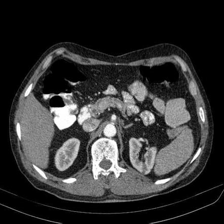
Celiac artery compression syndrome in children adolescents and young adults. Radiology plays an important role in the diagnosis of median arcuate ligament compression. Ischemia results from external compression of the proximal celiac artery during respiration typically during expiration by the median arcuate ligament just below the diaphragm. The definition of the syn-drome relies on a combination of both clinical and radiographic features. It typically occurs in young patients 2040 years of age and is more common in thin women who may present with. Median arcuate ligament MAL syndrome MALS also known as celiac artery CA compression syndrome and Dunbar syndrome occurs because of extraluminal compression of the CA root by the MAL which is part of the diaphragm. The aim of the paper is to determine the incidence of celiac artery compression CAC based on computed tomography CT scan and correlate the findings to the clinical presentation of patients presenting for CT scan in a hospital. It has a female to male ratio of 41. The median arcuate ligament syndrome or celiac artery compression syndrome was first described in 1963 by Harjola 3.
Keywords Key words celiac artery celiac artery compression syndrome CTA extrinsic compression. 1Department of Radiology AZ Sint-Maarten Duffel-Mechelen Belgium. The aim of the paper is to determine the incidence of celiac artery compression CAC based on computed tomography CT scan and correlate the findings to the clinical presentation of patients presenting for CT scan in a hospital. Celiac artery compression syndrome is a rare condition with a reported incidence of 2 per 100000 population. This latter is a fibrous arch connecting the right and left diaphragmatic crura on. Celiac artery compression syndrome. The compression of the proximal part of the celiac trunk by median arcuate ligament of the diaphragm during expiration is defined as median arcuate ligament syndrome.





















Post a Comment for "Celiac Artery Compression Syndrome Radiology"